A Comprehensive Guide To Atrial Septal Defects In The Heart

There's no question that the heart is one of the most vital organs of the body, and when something is awry with the heart it can make the difference between life and death. Although we often think of heart problems as occurring later in life, someone can experience heart problems at any stage of their lives, and a heart defect can even be present at birth, says the Centers for Disease Control and Prevention (CDC). One of the most common congenital heart defects is called an atrial septal defect, which affects approximately 25% of children, according to a 2022 article published in StatPearls.
For those who aren't familiar with the medical term, an atrial septal defect is a defect that can occur at birth where a hole is present between the upper chambers of the heart, as described by the Mayo Clinic. A child with an atrial septal defect may have incorrect blood flow as their blood travels across the hole and into the lung arteries (per American Heart Association). For this reason, having a larger defect can gradually damage the lungs because of how the heart and lungs work harder to handle the excess blood.
Many babies will not experience signs or symptoms when they are born with an atrial septal defect, and the defect can even be diagnosed in adulthood (per CDC). In severe cases, someone who is affected may have trouble breathing, frequent lung infections, and heart murmurs, and they could be at an increased risk of a stroke.
How are atrial septal defects evaluated and diagnosed?
It can be scary knowing that there's a condition that involves a hole in your heart, and learning more about atrial septal defects could make you wonder about how this defect is diagnosed. Initially, a child who is suspected to have an atrial septal defect may be referred to a pediatric cardiologist who specializes in treating children and adolescents with heart disease, as explained by KidsHealth. Diagnostic tests can then be conducted to determine whether the child was born with the defect.
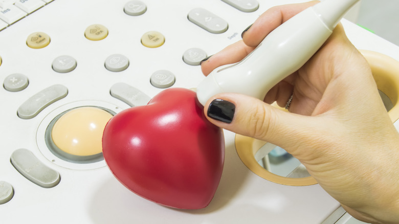
The most commonly used imaging test to diagnose an atrial septal defect is an echocardiogram, which uses sound waves to generate visual images of the heart's movements (per Mayo Clinic). According to a 2015 article published in Clinical Medicine Insights: Cardiology, echocardiography can be used to measure the size and shape of the hole in the heart, the directionality of blood flow, as well as search for any other abnormalities that may exist.
In addition to echocardiography, a doctor may recommend completing a chest X-ray, electrocardiogram (EKG), and computed tomography (CT) scan, as listed by the Mayo Clinic. If the results of echocardiography come back as inconclusive, a cardiac magnetic resonance imaging (MRI) scan may also be used to create more detailed images of the heart in hopes of clarifying the diagnosis.
What are the treatment options for atrial septal defects?

Once a formal diagnosis is made, how is an atrial septal defect treated? According to the Mayo Clinic, what avenue of treatment someone might need to take depends on the size of the hole in their heart. For some individuals, it's possible for their atrial septal defects to close on their own during childhood. Furthermore, some of the smaller defects may not require treatment if they aren't causing problems.
One of the less invasive treatment options for atrial septal defects is called cardiac catheterization, which involves the insertion of a catheter into a blood vessel in the groin area that is guided to the blood vessels in the chambers of the heart to create measurements, explains UCSF Health. Because the area is numbed before the catheter is inserted, cardiac catheterization is a generally painless procedure (per Hackensack Meridian Health). Once the size of the hole has been measured, a septal repair device can be inserted if the defect is small enough. Within three months of being placed, the hole in the heart will close as the lining of the heart wall forms over the patch.
If the size of the hole is too large to have cardiac catheterization, the American Heart Association reports that open-heart surgery may be necessary to close the hole. During open-heart surgery, a surgeon will create an incision in the chest wall and use patches to repair the hole in the heart (per Mayo Clinic).